The Michael J. Fox Foundation (MJFF) recently announced 142 new research grants awarded in December 2025 and January 2026 totaling $101 million to advance Parkinson’s disease (PD) research. These investments support projects that deepen our understanding of what drives PD, improve how we measure it and open new possibilities for treatment.
From cutting-edge brain imaging to precision genetic approaches to and new strategies to address freezing of gait, the funding is designed to move promising science forward for people living with Parkinson’s.
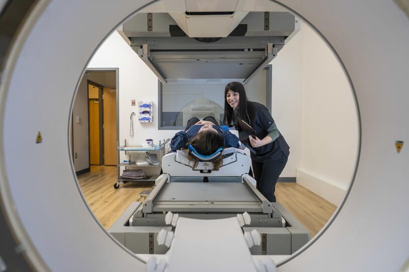
Making Parkinson’s Visible with Advanced Imaging
Seeing how Parkinson’s unfolds within a person’s brain helps researchers better understand its biology, improve diagnostic accuracy and evaluate whether new therapies are working. MJFF is investing in advanced imaging tools that make the underlying disease processes visible in real time, enabling more precise diagnosis and more effective tracking of treatment impact. This work is supported through initiatives such as the Parkinson’s Progression Markers Initiative (PPMI), the Foundation’s Biomarker Advancement Program, imaging consortia and targeted efforts to develop alpha-synuclein imaging agents — all designed to accelerate the discovery, validation and broad use of these critical tools.
A New Window into Brain Chemistry
One promising effort focuses on using metabolic MRI (a type of brain scan that detects chemical changes inside brain cells) to measure small molecules produced in the brain that fuel, regulate and maintain neuronal function. PD-linked inflammation and mitochondrial dysfunction (impairments in the cell’s energy-producing structures) can disrupt the functioning of these small molecules. By capturing their chemical signals, scientists hope to better distinguish between early, mid-stage and more advanced PD. This approach could help doctors more accurately track disease progress and response to potential treatments.
Measuring Inflammation in Real Time
Researchers are also developing a new MRI probe that highlights areas of inflammation in the brain. The probe, called Fe-PyC3A, becomes visible on a scan when it encounters certain oxidants that are signs of inflammatory activity. If validated, this tool could allow researchers to more easily detect, quantify and measure changes in brain inflammation in people with PD. Because chronic inflammation can damage dopamine-producing neurons, being able to measure it may help researchers better understand how it contributes to worsening symptoms. This tool could also aid in PD drug development by allowing researchers to see whether anti-inflammatory drugs are working early in a clinical trial so researchers can change tactics quickly if necessary.
Tracking Harmful Proteins
Parkinson’s and other neurodegenerative diseases are marked by a toxic buildup of abnormal proteins in the brain. While progress has been made in identifying some of these proteins in people with Alzheimer’s using PET scans, the ability to image these proteins in people with PD is still in development.
MJFF is supporting studies to identify PET imaging tracers — special compounds that bind to specific proteins and make them visible on a brain scan — that target alpha-synuclein as well as a specific form of tau known as 4R-tau. Measuring these specific proteins in the living brain is important because alpha-synuclein is a core driver of Parkinson’s pathology and 4R-tau is linked to related disorders that can resemble PD.
If successful, these tools would allow researchers to directly measure protein buildup in living patients, potentially improving diagnosis, help match patients to the right clinical trials and provide a faster way to see whether treatments are reducing toxic protein accumulation.
Targeting the Root Causes of PD
Parkinson’s affects people differently, and in some cases, specific genetic changes increase risk. Understanding these genetic contributors can help scientists develop new treatments and personalize PD care.
A major focus of MJFF’s longstanding research on the genetic causes and contributors to PD is LRRK2. In some people with PD, mutations in the LRRK2 gene cause its enzyme activity to become abnormally high, disrupting normal cellular cleanup and contributing to toxic protein buildup in the brain, making it an important target for research and therapeutic development. Through initiatives such as the LRRK2 Investigative Therapeutics Exchange (LITE), targeted funding programs within its genetic and biomarker portfolios and large-scale studies such as PPMI, MJFF is working to better understand how LRRK2-related Parkinson’s develops and progresses, helping ensure that promising treatments can be tested efficiently and in the right patient populations.
Confirming Drugs Are Working as Designed
Several therapies in development aim to reduce LRRK2 activity. To move these therapies forward, researchers need reliable ways to measure whether a drug is having the intended effect.
MJFF is funding the validation of tests that measure increases and decreases in LRRK2 (and an activated form of LRRK2 known as pS1292) in blood and other biofluids. These tests could help confirm that a therapy is working at a molecular level, information needed for drug approval. These tests also could help identify which patients are most likely to benefit from LRRK2-targeted treatments, helping advance a more precision medicine approach to PD treatment.
Defining Biological Signatures
Researchers are also studying how LRRK2 mutations affect the body beyond the gene itself. One such project is analyzing oxylipins — lipid molecules involved in inflammation and neurodegeneration — in people with and without LRRK2 mutations. If LRRK2 mutations are linked to specific oxylipin profiles, then these profiles could potentially help researchers track disease progression or identify subgroups of patients with LRRK2-associate biology changes.
Another study focused on LRKK2-related pathology is examining small clumps of alpha-synuclein found in people with LRRK2 mutations. In most patients, alpha-synuclein forms large clumps called Lewy bodies that can be seen under a microscope, but some patients with the LRRK2 gene often have smaller, less-visible protein clumps. Scientists are investigating whether the smaller protein clumps spread between cells and how that might contribute to disease progression.
Addressing Freezing of Gait
Freezing of gait (FOG) is one of the most disabling symptoms of Parkinson’s. During a freezing episode, a person may feel as though their feet are stuck to the floor. These unpredictable episodes can increase fall risk and significantly impact independence.
For many people, medication and traditional deep brain stimulation (DBS) do not fully relieve freezing. MJFF is investing in new strategies to better understand, measure and treat this challenging symptom. Through initiatives such as the Freezing of Gait (FOG) in Parkinson’s Disease Research Program, the Therapeutic Pipeline Program and other targeted grants, the Foundation is accelerating research to better define the biology of freezing, objectively track episodes and test innovative therapies designed specifically to address them.
Mapping the Brain Networks Behind Walking
In one funded project, researchers are working to better understand a brain network called the Somato-cognitive action network (SCAN), which may be involved in the motor symptoms seen in Parkinson’s. This recently identified brain network links parts of the brain involved in controlling the hands, feet and mouth.
Using advanced MRI and detailed gait testing, the researchers are studying how this network functions in people with Parkinson’s, both on and off medication. By linking brain connectivity changes to FOG, the findings could guide more precise brain stimulation strategies and identify this network as a target for these therapies.
Testing a New DBS Approach
Another funded project seeking to improve FOG by targeting a brain structure called the cuneiform nucleus with deep brain stimulation. The cuneiform nucleus plays a key role in starting and stopping walking, and a pilot study by the research team recently showed targeting it with DBS may help reduce FOG.
The researchers will use walking tests, wearable devices and brain activity recordings to measure changes in movement and brain function to assess the impact of targeting the cuneiform nucleus with DBS. If successful, this approach could open a new treatment option for patients with FOG who currently have limited alternatives.
Moving from Discovery to Better Care
Together, these research investments reflect MJFF’s comprehensive strategy to tackle Parkinson’s from multiple angles. By developing next-generation imaging and digital tools, researchers can measure disease more precisely and determine whether therapies are truly changing its course. By advancing genetic research, scientists are working toward treatments tailored to specific biological drivers. And by targeting freezing of gait, investigators are addressing a symptom that has a profound impact on daily life.
Each of these efforts strengthens the pipeline from scientific discovery to the development of therapeutics. Step by step, they bring us closer to treatments that slow or stop Parkinson’s disease and improve the quality of life for everyone living with it.
To learn more about active funding mechanisms at MJFF, visit our Funding Opportunities page.